The Duke University Health System (DUHS) is a regional provider of health care services.
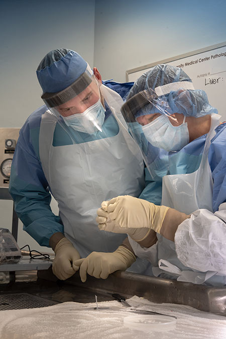
DUHS comprises three hospitals with 1,498 beds. Its affiliation with the Durham Veterans Affairs Hospital (274 beds) provides additional training opportunities. Duke laboratories perform more than 7 million laboratory tests per year; over 60,000 surgical pathology cases and 48,000 cytology cases, and over 350 autopsies.
In addition, DUHS provides a significant level of molecular pathology testing including flow cytometry, image analysis, molecular diagnostics, cytogenetics, etc. The Pathology Department is fully equipped with the most modern technology for diagnosis and research. The Seeley Mudd Library is also available located centrally in the medical complex. In addition, residents have 24-hour access to the Forbus Reading Room, which houses essential textbooks and ample study materials. Within the department, the residents have personal carrels and are provided with laptop computers and microscopes.
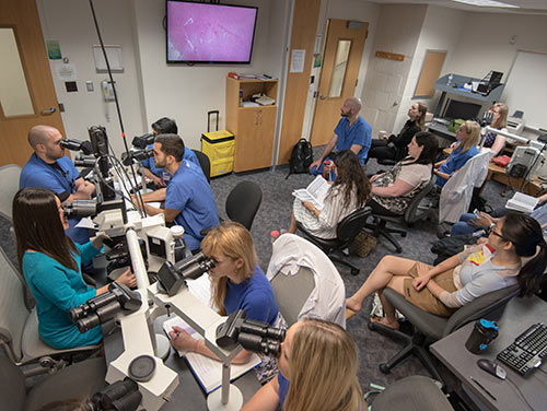
Our state-of-the-art media room allows for teaching and meetings in many configurations. Equipped with automated room controls, all computer and media functions, live-image microscope, and digital projector, the room is a versatile and comfortable space for departmental use.
Teaching in the Duke University School of Medicine
Excellence in teaching has been a hallmark of this department since its inception. All residents in the department teach medical students and assist the faculty in the Pathology Course teaching laboratories.

Durham VA Medical Center
Residency training in pathology at the Durham VA Medical Center (DVAMC) forms an integral part of the pathology residency training program at Duke University Medical Center. The DVAMC offers rotations in Surgical Pathology, Diagnostic Electron Microscopy, Blood Banking, Clinical Chemistry, Coagulation, and Microbiology, During these rotations, the residents work solely at the DVAMC under the direct supervision of VA staff pathologists. Residents on Autopsy Pathology and Immunopathology rotations participate in these services at Duke and the DVAMC simultaneously. On both services, work at the DVAMC is under the direct supervision of VA staff pathologists. Residents may also be assigned to the DVAMC for the Lab Management Apprenticeship.
Forensic Pathology
The State of North Carolina is fortunate in having a unified medical examiner system located in Raleigh, directed by the Chief Medical Examiner. All Duke pathology residents are required to spend one month in forensic pathology during their training program, usually in their third or fourth year.
Resources
Within the Department of Pathology, the residents have 24-hour access to the Forbus Library that houses up-to-date textbooks, current journals and ample study materials for board examinations. The residents also have access to the Duke Medical Center Library during normal working hours from both Duke and the Durham VA (directly across the street). Residents have access to on-line medical literature databases including ExpertPath, Immunoquery, and ASCP Question Bank and Path Primer 24 hours a day.

Conference Room
Our conference room allows for teaching and meetings in many configurations including virtual conferences. Equipped with automated room controls, all computer and media functions, live-image microscope, and digital projector, the room is a versatile and comfortable space for departmental use.
