
On Feb. 16th, 2024, Professor of Pathology Sara E. Miller, PhD, was invited to give a virtual presentation to the State Microscopical Society of Illinois as part of a teaching outreach program offered to local microscopy societies by the Microscopy Society of America (MSA), an international microscopy organization.
Miller established the Duke EM Virology Laboratory, which she directed for over three decades. She currently is director of the Center for Electron Microscopy and Nanoscale Technology, the biological ultrastructural imaging core at Duke. She has served as an MSA tour speaker many times in the past, traveling all over the United States, and has also served as both councilor and president of the MSA.
In her talk, titled “Identification of Viruses by Electron Microscopy (EM): Basic Techniques and Pitfalls,” she described methods for preparing specimens for examination by EM and characteristics of viruses used in their identification. In addition, she covered pitfalls in diagnosis and how to avoid them, a major one being that some normal cellular organelles can resemble viruses.
While there are more sensitive tests, EM has the advantage of not requiring special reagents to recognize viruses. Any immunological or molecular test requires a special "tag" that will bind to them or their components; this can be a problem if one doen't know what to look for. First, an a priori guess must be made as to which pathogen might be present, then the reagent (e.g., antibody, nucleic acid) with the corresponding binding site must be selected. If the wrong tag is chosen, the test will be negative, even though the patient may have a viral infection. Furthermore, reagents do not exist for all viruses. Here, different kinds of viruses are shown by two methods, negative staining and thin sectioning:
Legend:
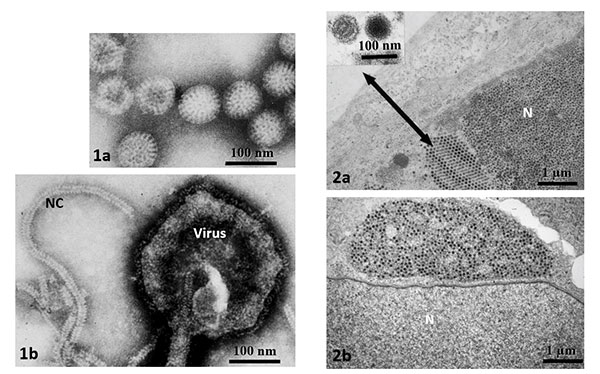
1. Negative stains of viruses from fluids placed on a support; the background is stained dark by a heavy metal salt solution.
a. Rotavirus, a naked icosahedral virus (causes diarrhea and vomiting).
b. Measles virus, an enveloped virus with spikes on the outside and a helical nucleocapsid (NC) (causes fever and rash).
2. Ultrathin (70 nm) sections of virus-infected tissues.
a. Adenovirus, a DNA virus constructed in the nucleus (N) (causes infections in many organs).
b. Reovirus, an RNA virus constructed in the cytoplasm––the clump of particles just above the nucleus (can cause respiratory infections; is a cousin to rotavirus).