![]()
The Duke Center for Electron Microscopy and Nanoscale Technology is a resource for high-quality, high-resolution light and electron micrographs. It’s the longest continuously running shared service facility on campus with a history of over four decades.
This laboratory provides a full range of sample preparation, technical assistance, and help in the design of experimental procedures, so that you can have the best images of your specimens to answer your scientific questions. We also can train researchers/students in all aspects of electron microscopy if desired. We supply ultrastructural analyses of small specimens, including cells and tissues; sub-cellular organelles; and microorganisms, such as viruses, bacteria, fungi, and parasites.
We also work on your selected images and prepare them for publication as requested by the journal. For new experiments by inexperienced users, we recommend that you consult the EM staff before starting a project to ensure proper planning of your study and harvesting of the specimens.
There is no charge for consults. Dr. Miller and Dr. Ferreira are both highly-experienced investigators who are able to comprehend your proposal and provide guidance on how electron microscopy can help answer questions or corroborate your findings.
Writing a grant? Ask us how EM can add weight to your proposal. We can also assist in writing the microscopy sections.
For information on use of the facility, capabilities, or specimen preparation, contact the director or manager:
saram@duke.edu
919 684-9141
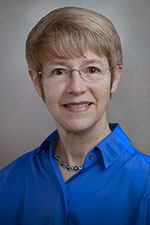
Dr. Miller attended Wake Forest University and the University of Georgia and did postdoctoral studies at the University of North Carolina at Chapel Hill. She has since served on the faculty at Duke Medical Center teaching microscopy, ultrastructure, and virology for several decades. Her double major in microbiology and chemistry provides the training for investigating the best methods for processing and analyzing myriad specimens. Her extensive career in electron microscopy has prepared her for leading ultrastructural studies on a wide variety of sample types, including viruses, bacteria and other microbes, plus numerous tissue types from many species. She was elected a Fellow of the Microscopy Society of America (MSA) and served as president of the MSA and president of the Society for Ultrastructural Pathology, both international academic organizations, as well as president of the Southeastern Microscopy Society.
davis.ferreira@duke.edu
919 684-3452
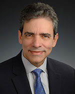
Dr. Ferreira has a PhD in microbiology from the Federal University of Rio de Janeiro, Brazil. Before joining our team, he was a virology professor at that institution for over 20 years. He studied as a postdoctoral fellow at North Carolina State University, where he worked as adjunct faculty and principal research scientist in the Department of Molecular and Structural Biochemistry. Dr. Ferreira also was a Director of Research and Development for Q2 Solutions (IQVIA) in the vaccines department in Durham, N.C. Having a propensity for research, he came back to academia, becoming the manager of the biological EM core at Duke in 2023. During his trajectory as researcher, faculty member, and industry laboratory director, he developed a unique multidisciplinary expertise and vision that prepared him to collaborate with investigators and guide research projects.
EM Core Capabilities
- Transmission Electron Microscopy (TEM)
- High-resolution digital EM imaging (16 MP camera)
- Ultra-microtomy (60-90 nm thin sections of cells and tissues)
- Ultra-cryomicrotomy (thin sections of cells and tissues for immuno-labeling)
- Negative staining of viruses and sub-cellular organelles
- Immuno-labeling of particulate specimens
- “Vibratome” (vibrating tissue slicer sectioning) sectioning
- Pre-embedding immuno-labeling
- Silver-enhanced small-tag immuno-labeling
- Microwave specimen processing
- High-resolution (12 MP camera) photo microscopy (dissecting and conventional light microscopy)
- Stereoscopy with darkfield imaging
- Digital image processing; stitching, extended focus, measurement
- Scanning Electron Microscopy (SEM)
- Critical point drying for SEM
- Metal coating for SEM
- Microprobe (X-ray) elemental analysis
For examples of sample types and micrographs, see our Gallery, and for services and pricing, see our User Fees at the click-on menus.
Location
Davison Building (Green Zone), Rooms 245M, 252M-E and 314M
Duke Clinics (Duke South)
40 Medicine Circle
Durham, NC 27710
Postal Address
DUMC 3712
Durham, NC 27710 USA
Phone: 919-684-3452
Guide for sample harvesting, handling, and fixing:
- Handle/harvest samples carefully so as not to mash them.
- Slice them with a scalpel; do not use scissors.
- Do not squeeze them.
- Do not pick them up with forceps.
- Lift them with a spatula or sable hair paint brush.
- Make sure that one dimension is only ~1 to 1.5 mm thick (a sliver).
- Length is irrelevant, but at least one dimension should be very thin for fixative infiltration.
- If a special orientation is needed (e.g., intestine, skin, kidney) call the lab for instructions.
- Put the slices into fixative in vials such as scintillation vials that have a big enough mouth to get a pipet into to change solutions and get the tissue out of without damaging it.
- Eppendorf tubes are too small. Besides not having enough volume, they're too small to work with, i.e., to change the solutions (>20 times).
- Use about 4-8 ml of fixative* per vial, depending on the numbers of pieces.
- Use a different vial for anything you want to keep separate from something else.
- Cells in suspension and monolayer cultures must be handled differently. Call the lab for instructions. (919 684-3452) We must have enough cells to see a pellet in a 0.5 ml tube (at least 1-2 million cells).
- Label vials with a permanent marker clearly (legibly) with a designation that matches the legend on the intake sheet (see QR Code).
- Put samples into some order that makes sense (e.g., 1, 2, 3, 4; A, B, C, D; WT, KO1, KO2, KO3; Control, Treated1, Treated2, Treated3; etc.; with the most normal first, followed by those with increasing amounts of changes expected.
- Do not ever freeze the samples, either before or after fixation. Use perfusion fixation for animal tissues if possible; if not, use immersion fixation after dissection.
- Do not put all the tissue into paraffin for light microscopy and then decide that you want EM. Yes, we can take it out of wax and embed it, but the ultrastructure will be compromised, and the fine structure in which you're interested may or not be evident.
- Deliver samples to the manager or director (call first) and bring the completed intake sheet or leave samples in the refrigerator in 252C Green Zone with your name clearly marked on the box or rack and the completed intake sheet pinned to the front of the refrigerator. Call or text to alert us that you have delivered them.
*Fixative
- The type of fixative used immediately after tissue or cell harvest will depend on the results desired. Immuno-labeling studies require a fixative with a composition different from conventional TEM. Consult the EM staff for advice.
- Call or email for instructions on fixatives for perfusion of animals.
- Particles, such as viruses in media for negative staining should not be fixed. Contact our staff before preparation and before delivery and be prepared to provide information on virus type and pathogenicity.
Solutions
- Routine initial fixative: 2-4% buffered glutaraldehyde
- Perfusion fixative: 2-4% buffered paraformaldehyde or 2-4% buffered paraformaldehyde plus 0.1-0.5% glutaraldehyde. Note: for immuno-labeling, the glutaraldehyde concentration should be kept low (0.05-0.5 %); call to discuss.
- For conventional TEM, osmium tetroxide is used after the aldehyde fixation for contrast; this step is performed in the EM lab.
- Buffer: 1 M cacodylate, pH 7.2-7.4 (best) or 1 M phosphate pH 7.2-7.4
Fees
External users, call 919-684-3452.